Diagnosis · Thyroidectomy · Sternotomy-Sparing Surgery · Endocrine Care · Recovery
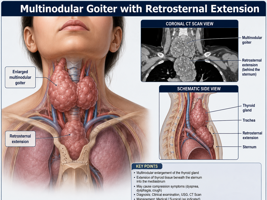
Multinodular Goiter (MNG) is the enlargement of the thyroid gland with multiple nodules. When the gland grows downward behind the breastbone (sternum) into the chest cavity, it is called retrosternal or substernal goiter. This condition can compress the windpipe, food pipe and major blood vessels, leading to breathing difficulty, swallowing problems and serious complications if left untreated.
India is a leading destination for retrosternal goiter surgery, offering advanced thyroidectomy, sternotomy-sparing techniques, intraoperative nerve monitoring (IONM) and minimally invasive endocrine surgery at JCI/NABH accredited centres — at a cost 60–80% lower than the USA, UK or Europe, with outcomes that match international benchmarks.
The thyroid gland is a butterfly-shaped organ located in the front of the neck that produces hormones regulating metabolism. A multinodular goiter develops when multiple lumps (nodules) form within the gland, causing it to enlarge. Over years, the enlarged gland can extend downward through the thoracic inlet into the chest, behind the sternum — this is called retrosternal extension.
Retrosternal goiters are clinically significant because the narrow space behind the sternum is shared with the trachea (windpipe), oesophagus (food pipe), recurrent laryngeal nerves and great vessels. As the goiter grows, it compresses these structures and causes obstructive symptoms. Approximately 5–15% of all goiters have a retrosternal component.
Substernal/Retrosternal Goiter — more than 50% of the gland lies below the thoracic inlet. Intrathoracic/Mediastinal Goiter — extends deep into the mediastinum, often requiring sternotomy. Cervico-Mediastinal Goiter — partly in the neck, partly in the chest.
Primary (true) retrosternal goiter — rare; develops from ectopic thyroid tissue with its own blood supply from intrathoracic vessels. Secondary retrosternal goiter — common; downward extension of a cervical goiter through the thoracic inlet, with blood supply from the inferior thyroid artery.
Euthyroid (normal function) — most common. Toxic MNG (Plummer's disease) — autonomous nodules producing excess hormone. Hypothyroid MNG — less common, usually associated with iodine deficiency or autoimmune thyroiditis.
The risk of cancer in retrosternal MNG is 3–17%, similar to or slightly higher than cervical MNG. Any suspicious nodule must be evaluated with FNAC before surgery.
Symptoms develop gradually as the goiter enlarges and compresses surrounding structures. Many patients are asymptomatic for years before presenting with obstructive features.
A combination of clinical examination, blood tests and detailed imaging is required to confirm the diagnosis, define the extent of retrosternal extension and plan surgery.
Surgery is the treatment of choice for retrosternal MNG due to the risk of progressive airway compromise, swallowing difficulty and underlying malignancy. Indian centres offer the full spectrum of modern endocrine surgical care.
Complete removal of the entire thyroid gland is the procedure of choice for retrosternal MNG. It eliminates the risk of recurrence, allows complete histopathological examination, and prevents future malignancy. Performed through a standard cervical (Kocher) incision in 95–99% of cases, even for large retrosternal extensions. The retrosternal portion is usually delivered through the neck incision using gentle blunt mobilisation.
Only 1–5% of retrosternal goiters require partial or full sternotomy. Indications include: deep mediastinal extension below the aortic arch, primary intrathoracic goiter with independent blood supply, suspected malignancy with invasion, recurrent retrosternal goiter, or massive size with significant vascular displacement. Indian centres routinely perform partial manubriotomy (limited bone cut) to minimise morbidity and recovery time.
Modern Indian endocrine surgery centres use continuous intraoperative nerve monitoring of the recurrent laryngeal and superior laryngeal nerves to minimise the risk of vocal cord paralysis. This is particularly valuable in retrosternal cases where the nerve anatomy may be distorted or stretched by the goiter.
Use of advanced energy devices — Harmonic Scalpel, LigaSure and bipolar diathermy — reduces blood loss, operative time and post-operative hematoma risk. For purely cervical MNG, minimally invasive video-assisted thyroidectomy (MIVAT) and robotic transaxillary thyroidectomy may be options, though retrosternal extension usually requires open conventional surgery.
RAI ablation has a very limited role in retrosternal MNG because the gland is large and may not respond well to iodine uptake. It is generally reserved for patients with toxic MNG who are unfit for surgery or refuse it. RAI does not reliably relieve compressive symptoms and is not a substitute for surgery in symptomatic retrosternal disease.
Most patients are discharged within 2–4 days after a cervical thyroidectomy and 5–7 days after a sternotomy. Recovery milestones include:
Indian thyroid surgery centres perform 1,000+ thyroidectomies per year with retrosternal case experience.
MS/MCh-qualified surgeons trained in dedicated endocrine and head-neck surgery fellowships globally.
Retrosternal thyroidectomy with IONM and ICU stay at a fraction of Western costs.
Surgery scheduled within 1–2 weeks of arrival — no months-long NHS-style queues.
* Costs vary by hospital, surgeon experience, length of stay, ICU requirement and use of IONM. Contact Satyug Healthcare for a personalised estimate.
The prognosis after total thyroidectomy for benign retrosternal MNG is excellent. In experienced hands, mortality is under 1%, recurrent laryngeal nerve injury under 2%, and permanent hypoparathyroidism under 2–3%. Compressive symptoms — breathing difficulty, swallowing problems and stridor — resolve dramatically within days. With lifelong levothyroxine replacement and periodic TSH monitoring, patients return to a completely normal quality of life. If malignancy is found on histopathology, the prognosis depends on the type and stage but is generally favourable for well-differentiated thyroid cancers (95%+ 10-year survival).
Travelling abroad for retrosternal goiter surgery needs careful coordination — from imaging review and surgeon selection to visa, hospital admission and post-op follow-up. Satyug Healthcare manages every step.
Send TSH, T3, T4, ultrasound, CT scan, FNAC and laryngoscopy reports — get a written opinion from a leading endocrine surgeon.
Consultations with two or more endocrine and head-neck surgeons so you can choose who you trust most.
BLK-Max, Indraprastha Apollo, Medanta Medicity, Fortis Memorial, Max Saket, Manipal Dwarka, Artemis, Sir Ganga Ram Hospital, CMC Vellore, Tata Memorial.
Expedited medical visa invitation letter for patient and accompanying family members.
Airport pickup, accommodation, pre-op investigations, surgery scheduling, ICU and post-discharge care.
English, Arabic, Russian, French, Bengali — no communication barriers.
Video consultations to monitor TSH levels, thyroxine dose adjustment and surgical recovery.
Q1. Will I need a sternotomy (chest bone cut) for my retrosternal goiter?
No, in most cases. Over 95% of retrosternal goiters can be removed through a standard neck incision alone. Sternotomy or partial manubriotomy is needed only in 1–5% of cases — typically for deep mediastinal extension, primary intrathoracic goiter, recurrent disease, or suspected invasive malignancy. Your surgeon will decide based on your CT scan findings.
Q2. Will I lose my voice after surgery?
Permanent vocal cord paralysis is rare — under 2% in experienced hands, especially when intraoperative nerve monitoring (IONM) is used. Temporary hoarseness can occur in 5–10% of patients and usually resolves within weeks. Indian endocrine surgery centres routinely use IONM to minimise this risk.
Q3. Will I need lifelong medication after total thyroidectomy?
Yes. After total thyroidectomy, you will need lifelong levothyroxine (a once-daily tablet) to replace the hormones your thyroid would have produced. The dose is adjusted based on your TSH levels at 6 weeks and 3 months post-op, and once stable requires only annual monitoring. This is a simple, inexpensive medication with no major side effects when properly dosed.
Q4. What documents do I need to share to get a medical opinion?
Send: (1) recent TSH, free T3, free T4 reports, (2) anti-TPO and anti-Tg if available, (3) neck ultrasound report and images, (4) CT scan of neck and chest (most important — shows retrosternal extent), (5) FNAC report if done, (6) indirect laryngoscopy report, (7) ECG, ECHO and pulmonary function tests in elderly patients, (8) prior hospital records. WhatsApp or email — opinion within 24–48 hours.
Q5. How long will I need to stay in India for the surgery?
A typical timeline is: 3–5 days for pre-op consultation and investigations, 3–5 days hospital stay for cervical thyroidectomy (or 5–8 days if sternotomy is needed), and 5–7 days outpatient follow-up for suture removal and initial dose adjustment. Most international patients plan a 2–3 week stay. Affordable accommodation near the hospital is available for accompanying family.
Get a free written medical opinion from a leading endocrine surgeon in India within 24–48 hours — at no obligation.
📞 +91-8860606766 | +91-9910655125
✉️ query@satyughealthcare.com · 💬 WhatsApp 24/7