Medical Visa
Assistance
Assistance
Tele-Video
Consultation
Consultation
Language Interpretation
Services
Services
Second Medical
Opinion
Opinion
Post-Treatment
Care Coordination
Care Coordination
Coordinating care at accredited hospitals, including
Apollo HospitalsFortis HealthcareManipal HospitalsSarvodaya Hospital
Not sure where to start? Get a free medical opinion Not sure what your treatment will cost? Free, instant, itemised estimate -- treatment, visa, flights, hotel & food
What will your treatment cost?
Satyug Assistant
Online · usually replies instantly
Answers are informational, not medical advice.
Best Surgeon for Hemangioblastoma Surgery in India
Released Date: 2021-10-28
Written & reviewed by Vikram Kashyap, Medical Consultant, Satyug Healthcare
·Last updated: ·Editorial policy

🖼️ Understand It in Pictures
💡 In simple words
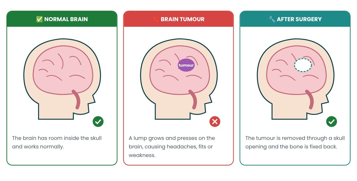
A brain tumour is a lump of abnormal cells that can press on the brain. In a craniotomy, the surgeon opens a small window in the skull, removes as much tumour as is safe, then puts the bone back.
Normal brain vs brain with a tumour
👀 Symptoms in Pictures

🩻 Scans & Tests — What Doctors See


🎬 The Treatment Journey in Pictures

📅 Recovery at a Glance
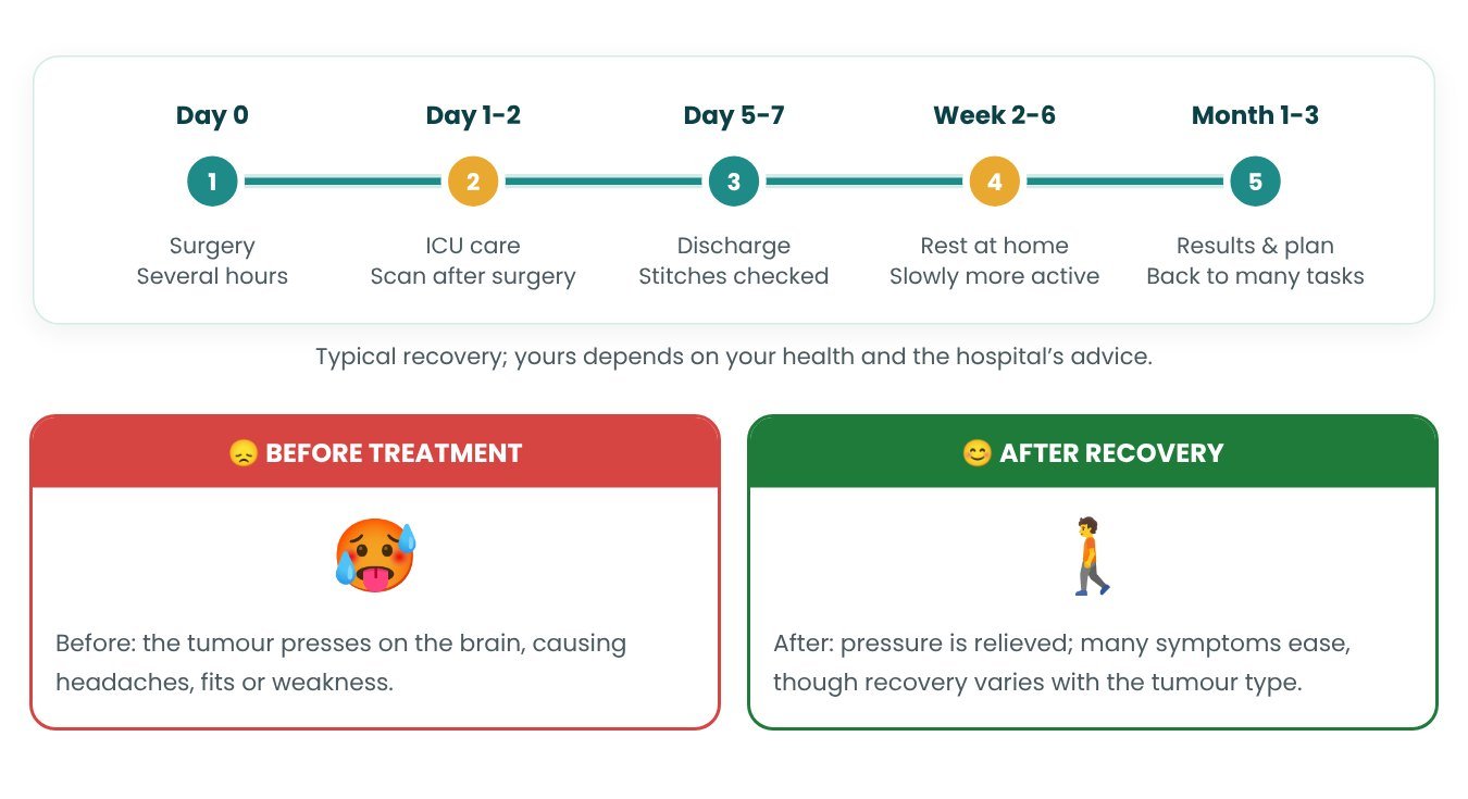
Drawings: Satyug Healthcare (simplified, not to scale). Photos: Wikimedia Commons; author and licence under each photo.
Hemangioblastoma is a benign, slow-growing tumor inside the brainstem or spine. It is a well-defined growth that forms in the lining of your blood vessels. A hemangioblastoma does not invade the surrounding healthy tissue and can be treated with surgery or targeted radiation.
Hemangioblastomas develop in the blood vessels of the brain, spinal cord and retina. These tumors can be caught early and removed before they become too large. The tumors are benign, which means they will not become cancerous. But they can put pressure on areas of the brain and cause various neurological symptoms. A hemangioblastoma often grows without an obvious cause. Recurrent hemangioblastoma may also suggest an underlying genetic condition such as von Hippel-Lindau (VHL) disease.
VHL can result in a hemangioblastoma. It is an inherited genetic condition. About 20% to 25% of hemangioblastoma cases are caused by VHL (von Hippel–Lindau disease). It affects 1 in 36,000 people.
VHL (von Hippel–Lindau disease) is caused by a gene mutation. This leads to the growth of benign tumors in many different parts of the body. The condition is diagnosed at an early age. There is no cure or prevention for VHL. The main treatment is surgical removal of the tumor. Hemangioblastomas often recur in people with VHL. They are the cause of death for 82% of people in that group.
Symptoms of a Hemangioblastoma
-
Headache
-
Loss of coordination
-
Imbalance
-
Nausea
-
Vomiting
Pressure on the brainstem and spinal cord can also cause:
-
Loss of motor function
-
Muscle weakness
-
Numbness in your extremities
-
Impaired bowel function
-
Impaired bladder function
Hemangioblastoma Diagnosis
Computed tomography (CT) scan.
Magnetic resonance imaging (MRI)
Cerebral angiography.
Hemangioblastoma Treatment
Surgery is the standard treatment option for a hemangioblastoma. This usually cures the condition unless the whole tumor cannot be removed. Your doctor may also use some of the diagnostic methods mentioned above to monitor your recovery after surgery.
There is a chance that surgery can remove the problem completely for people with VHL. Only about 25% of people receiving surgery for a hemangioblastoma ever require another surgical treatment.
Stereotactic radiosurgery (SRS). This form of radiation therapy can be used if surgery may lead to complications. It uses a focused dose of radiation to destroy the hemangioblastoma.
The beams of radiation stop blood flow to the tumor. This causes the tumor to shrink over several months. The aim of the procedure is to prevent the tumor from growing.
Some Important questions and answers:
What is VHL?
• Von Hippel-Lindau disease (VHL) is a multisystem neoplastic disorder that requires a multidisciplinary approach for optimal patient management.
What do VHL sufferers need?
• VHL patients require specific perioperative management of conditions such as pheochromocytomas, diabetes and renal insufficiency.
What do hemangioblastomas represent?
• Hemangioblastomas occur throughout the craniospinal axis in VHL patients. They show a saltatory growth pattern. Tumor edema precedes the development of peritumoral cysts, which is usually a precursor to symptomatic presentation.
Is surgical removal of hemangioblastoma safe?
• Currently, surgical resection of hemangioblastoma in VHL patients is usually reserved for symptomatic lesions. In most cases, surgical removal of CNS hemangioblastoma can be done safely and is effective and long-lasting.
How is hemangioblastoma removed?
• Hemangioblastoma resection is directed at the tumor, without resection, fenestration, shunting or other manipulation of the peritumoral cyst. Peritumoral cysts resolve after resection of the hemangioblastoma.
How can endolymphatic sac tumors present in VHL patients?
• Endolymphatic sac tumors (ELSTs) may present clinically as radiographic lesions in the petrous temporal bone of VHL patients, or with hearing loss or other vestibulocochlear symptoms. Hearing loss may be sudden or gradual in onset. Deafness results from tumor-related intralabyrinthine hemorrhage, tumor invasion of the otic capsule, or endolymphatic hydrops.
What sort of test does a VHL patient require?
• A detailed high-resolution radiographic examination is required in a VHL patient with vestibulocochlear complaints.
When is surgery done for ELST in VHL patients?
• Surgical resection is done in VHL patients with radiographically visible ELST to preserve hearing. Surgery can be considered in patients without radiographically evident ELST who have hearing loss and indirect evidence of ELST, e.g. intralabyrinthine hemorrhage.
We hope you have found some help in understanding this. Please consult your doctor for any other questions or help related to this. You can contact us anytime to get doctor's advice.
If you have any related query, you can send us your report at the email address below, or call or WhatsApp us for a second opinion.
Video/tele consultation also available
E-mail address- query@satyughealthcare.com
Phone number- +91888-242-4372, +91991-065-5125
Related Blogs





HAPPY PATIENTS
What Our Patients Are Saying






























Our Gallery