| Surgery Name | Cost | Room | Hospitalization |
|---|---|---|---|
| Chiari malformation treatment in India | Evaluation on OPD Basis- $800-$1000, Surgery-$7100 | single | Patient - 4 days in a room and 1 day in ICU |
1.Overstay more than package days,
2. Any other Specialty Consultations,
3. Special Equipment,
4. Additional Procedure/Surgery.
5. Blood Components.
🇮🇳 Medical Package — India
World-class treatment • NABH & JCI Accredited Hospitals • Save up to 80% vs USA/UK
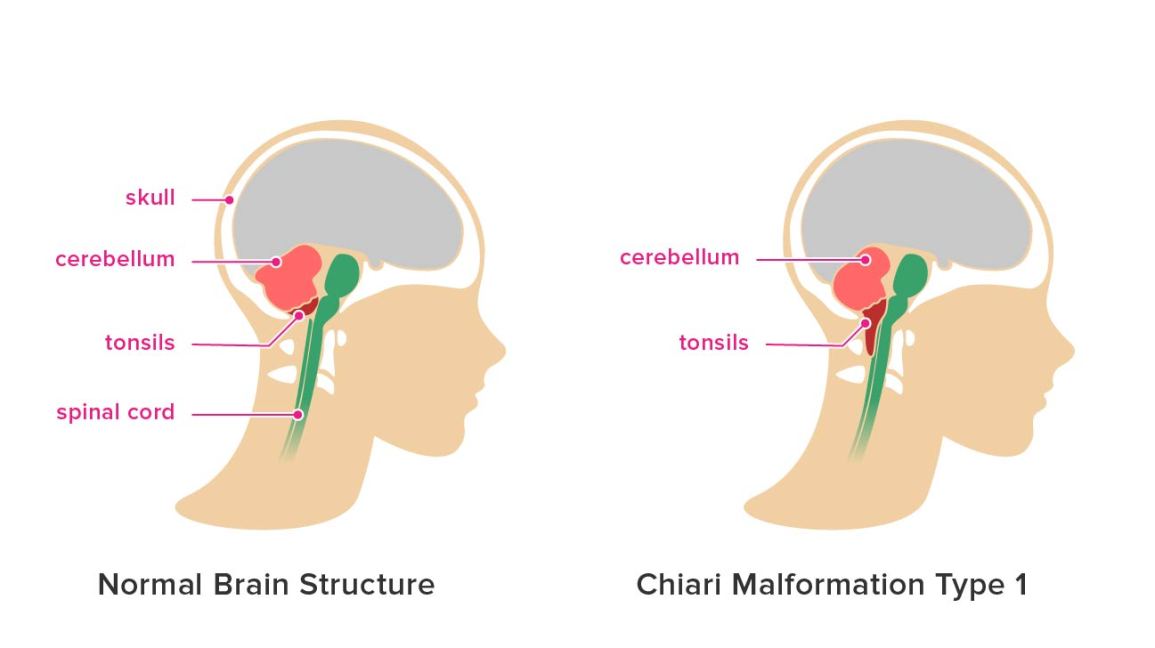
At the base of your skull, there’s an oval-shaped opening where the spinal cord is linked to the brain. This opening is known as the foramen magnum. A foramen magnum decompression is a procedure used to treat a condition called Chiari malformation, where the lower part of the brain at the skull base (cerebellum) extends into the spinal cord. Chiari malformation is a condition in which brain tissue extends into your spinal canal. It occurs when part of your skull is abnormally small or misshapen, pressing on your brain and forcing it downward.
The more common types of Chiari malformation are:
Type I
Type II
Chiari malformation type I
In Chiari malformation type I, signs and symptoms usually appear during late childhood or adulthood.
Headaches, often severe, are the classic symptom of Chiari malformation. They generally occur after sudden coughing, sneezing or straining. People with Chiari malformation type I can also experience:
Less often, people with Chiari malformation may experience:
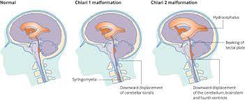
Chiari malformation type II
In Chiari malformation type II, a greater amount of tissue extends into the spinal canal compared with Chiari malformation type I. The signs and symptoms can include those related to a form of spina bifida called myelomeningocele that nearly always accompanies Chiari malformation type II. In myelomeningocele, the backbone and the spinal canal haven't closed properly before birth.
Signs and symptoms may include:
Chiari malformation type II is usually noted with ultrasound during pregnancy. It may also be diagnosed after birth or in early infancy.
Chiari malformation type III
In one of the most severe types of the condition, Chiari malformation type III, a portion of the lower back part of the brain (cerebellum) or the brainstem extends through an abnormal opening in the back of the skull. This form of Chiari malformation is diagnosed at birth or with an ultrasound during pregnancy. This type of Chiari malformation has a higher mortality rate and may also cause neurological problems.
When to see a doctor Symptoms
Magnetic resonance imaging (MRI). An MRI is often used to diagnose Chiari malformation. An MRI uses powerful radio waves and magnets to create a detailed view of your body. This safe, painless test produces detailed 3D images of structural abnormalities in your brain that may be contributing to your symptoms. It can also provide images of your cerebellum and determine whether it extends into your spinal canal. An MRI can be repeated over time, and it can be used to monitor the progression of your disorder.
Computerized tomography (CT) scan. Your doctor may recommend other imaging techniques such as a CT scan. A CT scan uses X-rays to obtain cross-sectional images of your body. A CT scan can help to reveal brain tumors, brain damage, bone and blood vessel abnormalities, and other conditions.
Treatment for Chiari malformation depends on the severity and the characteristics of your condition. If you have no symptoms, your doctor likely will recommend no treatment other than monitoring with regular examinations and MRIs. When headaches or other types of pain are the primary symptom, your doctor may recommend pain medication.
Doctors usually treat symptomatic Chiari malformation with surgery. The goal is to stop the progression of changes in the anatomy of your brain and spinal canal, as well as ease or stabilize your symptoms. When successful, surgery can reduce pressure on your cerebellum and spinal cord, and restore the normal flow of spinal fluid. In the most common surgery for Chiari malformation, called Foramen Magnum Decompression, your surgeon removes a small section of bone in the back of your skull, relieving pressure by giving your brain more room.
In many cases, the covering of your brain, called the dura mater, may be opened. Also, a patch may be sewn in place to enlarge the covering and provide more room for your brain. This patch may be an artificial material, or it could be tissue harvested from another part of your body. Your doctor may also remove a small portion of the spinal column to relieve pressure on your spinal cord and allow more space for the spinal cord. The surgical technique may vary, depending on whether a fluid-filled cavity (syrinx) is present, or if you have fluid in your brain (hydrocephalus). If you have a syrinx or hydrocephalus, you may need a tube (shunt) to drain the excess fluid.
Surgery involves risks, including the possibility of infection, fluid in your brain, cerebrospinal fluid leakage or problems with wound healing. The surgery reduces symptoms in most people, but if nerve injury in the spinal canal has already occurred, this procedure won't reverse the damage. After the surgery, you'll need regular follow-up examinations with your doctor, including periodic imaging tests to assess the outcome of surgery and the flow of cerebrospinal fluid.
If you have any related query, you can send us your report on below mention email address or you can call us or whatsApp for any second opinion.
Video/tele consultation also available.
E-mail address- query@satyughealthcare.com
Phone number- +91888-242-4372, +91991-065-5125
Free Medical Opinion
Expert review in 48 hours
Best Surgeon
Top specialists in India
Hospital Booking
NABH / JCI accredited
Visa Assistance
Invitation letter provided
Airport Transfer
Safe pickup & drop
Tele-Consultation
Video consult before travel
Hotel Booking
Near hospital, affordable
Multilingual Support
Arabic, French, English
📞 Get Started Today — It Is Free
Send your medical reports and get a free expert opinion within 48 hours. Our coordinators speak Arabic, French and English.
The most common symptom associated with a Chiari malformation is occipital headaches. These headaches are felt near the base of the skull and may radiate to cause pain in the neck and shoulders. They can be severe and may be described as sharp, brief, throbbing or pulsating.